COVID-19 causes brain damage.
It seems rather stark to see it written like that, without qualifications. But that sentence simply says something scientists have known for years.
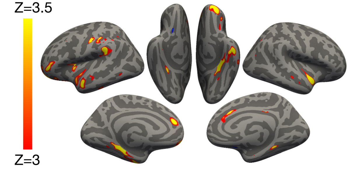
A watershed paper was published in Nature back in March 2022, showing definitively that SARS-CoV-2 (the virus that causes Covid) shrinks brain tissue.1 The researchers, Douaud and colleagues, used the UK Biobank of stored biological information and utilized the profiles of 384 subjects. They matched each of these subjects to controls (matched for age, socioeconomic factors, smoking status, etc.) that were negative for Covid. Not only that, they also had MRIs of those subjects — before subjects contracted the virus, and after.
It’s an elegantly designed study. Short of unethical testing (where the virus would be given to patients deliberately and then they would be scanned), this is the best data we have that shows an almost certain cause and effect. The virus causes brain damage.
New study
In the last month, another article has been published reinforcing this fact. Nature Medicine published the paper, but it is behind a paywall.2 However, an earlier version of the results was made available in January 2024 at Research Square.3 It is from that version that I quote the findings.4
These researchers, Greta Wood and colleagues, studied subjects who had neurological symptoms in the wake of SARS-CoV-2 infection, matching them against community controls at approximately an 1:8 ratio. There was also a non-neurological COVID cohort. The researchers found:
Prior to COVID-19 illness 11/137 (8%) NeuroCOVID and 15/152 (10%) COVID patients were concerned about their memory, increasing to 84/139 (60%) and 66/150 (44%) after COVID-19 illness respectively, of whom 35/82 (43%) and 45/66 (68%), respectively, perceived their memory problems to be progressive.5
What this means is, of those with a neurological component to their post-Covid condition, their concern about cognitive issues increased by more than sevenfold, and those who did not have a demonstrable neurological component to their post-Covid status still reported a fourfold increase in subjective memory problems.
For those who felt those problems were “progressive,” that indicates they perceived their issues to be worsening over time.
Analysis of individual tasks identified global impairment across all cognitive domains in both accuracy and response time (RT) in all clinical diagnostic groups [...] and no evidence for domain-specific deficits.6
This means that the impairment the researchers measured in these individuals was due to a global deficit. That is to say, the results did not indicate a focal problem, such as only numerical use or only language use, but rather a deficit of executive function in all categories that were included in the assessment.
Additionally, the respondents had consistently slowed reaction times, demonstrating what colloquially would be called dullness, a lack of being quick on the draw.
Compared to healthy controls, median [IQR] serum neurofilament light chain (Nfl-L, a marker of axonal injury), and glial fibrillary acidic protein (GFAP; a marker of astrocyte injury) were significantly raised in patients who had had COVID-19[,] and further raised in those with neurological complications[,] respectively[.] Tau was raised exclusively in those with neurological complications[.]7
Elevated levels of these proteins — Nfl-L, GFAP, and tau — are found in neurodegenerative diseases, such as Alzheimer’s disease.
In Alzheimer’s, the first aberrant protein that accumulates is amyloid-beta, yet a patient can live for decades in such a state with only minimal or mild symptoms. It is when tau accumulates and spreads, especially in a form called phosphorylated tau, is when Alzheimer’s becomes debilitating. Many experts have raised concerns that such markers manifest at such an early stage following SARS-CoV-2 infection.
Global volume composite had significant correlations with cognitive deficits in the overall cohort … The bilateral volume of anterior cingulate cortex was significantly and moderately positively correlated with overall cognition in the NeuroCOVID group[,] the COVID group[,] and the overall cohort[.]8
This indicates that the differences seen in brain volume between individuals was specifically linked to demonstrated cognitive deficits. One particular brain region of interest (ROI), the anterior cingulate cortex (ACC), was significant for this measure across all persons evaluated.
The ACC is a major hub of the brain, involved in several distinct functions. Those include emotion processing, pain perception, decision-making, anticipation of reward, resolution of conflicting information, and internal monitoring of autonomic data such as heart rate.
Subjective memory impairment was associated with inaccurate [...] and slow [...] responses on memory tasks and reduced superior temporal gyrus [...] and insula [...] volume. Raised Nfl-L in serum was weakly correlated with reduced thickness composite [...] and reduced superior temporal gyrus volume [...] and thickness[.]9
The superior temporal gyrus (STG) and the insula are two more ROIs that are important. The STG aids in auditory processing, especially that of language, and serves as a highway between the prefrontal cortex (the seat of higher cognition) and the amygdala (the “fear center”).
The insula, a node of grey matter deep beneath the surface of the brain, is involved in such varied aspects as interoception (that is, internal monitoring), emotion (especially in the interpretation of disgust and other “gustatory” emotions), empathy, visceral sensation, audition, and awareness of pain.
The scientists conclude:
This prospective, national, multicentre study of 351 COVID-19 patients who required hospitalisation with and without new neurological complications demonstrated that post-acute cognitive deficits, relative to 2,927 matched controls, were associated with elevated brain injury markers in serum and reduced grey matter volume. In contrast to studies early in the pandemic that identified dysexecutive syndromes predominant in acute infection[,] our study found global, persistent cognitive deficits even in those without clinical neurological complications. [...]
We have additionally shown that persistently raised serum GFAP was associated with post-acute cognitive impairment. GFAP is expressed by astrocytes, which participate in neuroimmune interactions within the brain. Its appearance in the plasma typically indicates injury of these cells and has been proposed as a prognostic biomarker for cognitive decline in the general population. [...]
Cognitive deficits were global, of significant magnitude, and spanned both accuracy and RT. Deficits were moderately to strongly associated with symptoms of depression, and the anterior cingulate cortex volume, which has functional roles in connecting cognition, attention, and emotion[.] … In our unsupervised cluster analysis, reduced cortical thickness, particularly in the superior temporal gyrus, was found to be associated with raised Nfl-L, potentially indicating a regional substrate for axonal injury in this population. [...]
Taken together, this prospective multicentre longitudinal cohort study found evidence of pervasive global cognitive impairment, associated with persistently raised brain injury markers, depression symptomatology, and reduced anterior cingulate cortex volume.10
For more on this study, please see Dr. Eric Topol’s Substack Ground Truths, where he posted about this in September.
The context
Now, I plan to follow this explanatory report with more speculative interpretations. I’m clearly separating them from what I’ve presented here, because I want readers to understand that what I’m presenting here is completely factual. But one thing that I want to impress before leaving off here is that preliminary data indicate that multiple infections of SARS-CoV-2 can compound effects. Some people may contract Covid once or twice without apparent complications but may then go on to observe long-term, lingering symptoms (called sequelae) after that third, fourth or fifth bout.
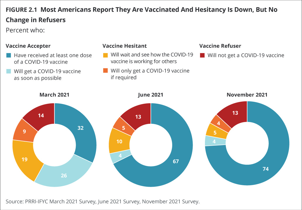
I mention this because we know that, from the start, there was an ideological divide as to the nature of vaccination as well as the simple acknowledgement that Covid was real. From the very beginning, far-right and ultra-libertarian folks refused to get vaccinated. This amounted to ~ 18-20% of the American adult population. (Many did not even want to mask!)
Unfortunately, this “ozone hole” of vaccination meant that we never achieved herd immunity, ensuring that Covid would continue to menace us, as indeed it has.
This ideological divide is important, because those are the people who are most likely to suffer multiple bouts of Covid contracture. Not only do they themselves tend to be unvaccinated, they tend to commingle with others who also are unvaccinated. Their chances of being exposed to the virus are higher than chance.11
Those who have had multiple bouts of Covid are more likely, over time and exposure, to manifest these more serious symptoms, particularly in this context neurological symptoms. Thus, it’s important for us to recognize that those persons may be more likely to exhibit certain behaviors linked to those symptoms. I’ll be exploring that more thoroughly under separate cover.
Gwenaëlle Douaud et al., “SARS-CoV-2 is associated with changes in brain structure in UK Biobank,” Nature (2022), Vol. 604, pp. 697-707.
Greta Wood et al., “Post-hospitalisation COVID-19 cognitive deficits at one year are global and associated with elevated brain injury markers and grey matter volume reduction,” Nature Medicine (2024).
Greta Wood et al., “Post-COVID cognitive deficits at one year are global and associated with elevated brain injury markers and grey matter volume reduction: national prospective study,” Research Square (2024).
It’s important to note that these findings were based on patients who were hospitalized with Covid. That’s a very specific segment of the population. But I want to stress here that cognitive dysfunction with Covid has been documented even in those who had a “mild” bout of sickness. In fact, the researchers found that, “[c]ompared to the COVID group, the NeuroCOVID group were more likely to have mild COVID-19” (ibid., p. 6).
Ibid., p. 6.
Ibid., p. 7.
Ibid., p. 8. (An axon is the part of a neuron that resembles a tail. It’s what the neuron uses to send information.)
Ibid., p. 8.
Ibid., pp. 8-9. (I’ve redacted the correlation coefficients (r values) for readability. I encourage those interested to read the paper in its entirety.)
Ibid., pp. 9, 10. Emphases added.
Additionally, some of these folks are the ones who disbelieved Covid’s existence altogether, so even if they came down with Covid since its inception, they almost certainly attributed it to some other disease (e.g., cold, flu). These are prime deniers who often have motivated reasoning to discount the evidence they’ve observed.


I didn't get COVID until after I'd been vaccinated. I know scores of other people in the same boat. I cannot say it's universal, but in my personal sphere, almost everyone I know who did get COVID were, in fact, vaxxed.
Do these studies differentiate between those who were vaccinated and non-vaccinated and acquired COVID?